
| Cardiac | |
|---|---|
| GI | |
| Bone | |
| GU | |
| Neuro | |
| Peds | |
| Faculty | |
| Student | |
| Quizzes | |
| Image DDX | |
| Mobile | |
| |
Misc |
| Videocasts | |
 |
LearningRadiology Imaging Signs
on Twitter
![]()
![]() Case of the Week 381
Case of the Week 381 ![]()
What sign is depicted on this image?
- 46 year-old with back pain
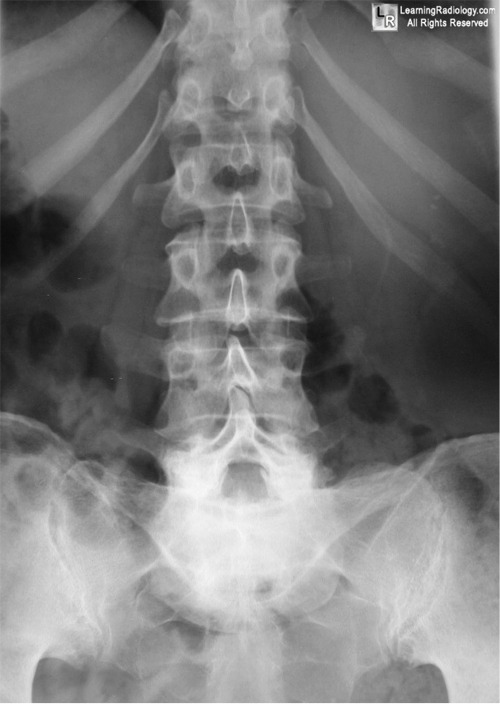
Frontal radiograph of lumbar spine
1. Dense pedicle sign
2. Ivory vertebra
3. Sherlock Holmes sign
4. Spine sign
5. Napoleon hat sign
Additional Images - Lateral radiograph of lumbosacral junction
![]()
Additional Images
.
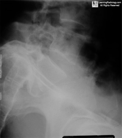
Lateral radiograph of lumbosacral junction
![]()
Answer:
.
5. Napoleon Hat Sign
.
.
More (Click Discussion Tab)
Napoleon Hat Sign
General Considerations
- Occurs with marked anterolisthesis of L5 on S1
- Visible on frontal radiograph of lumbar spine
- Implies the presence of bilateral spondylolysis and significant spondylolisthesis
- Spondylolysis resulting in this degree of spondylolisthesis is more often congenital and/or traumatic in origin and less often degenerative
- "Brim" of hat is formed by the downward rotation of the transverse processes
- "Dome" of hat is body of L5
- The sign can also be seen with marked exaggeration of the normal lordosis at the lumbosacral junction
.
This Week
46 year-old with back pain |
Part 2 reviews 50 more imaging signs, offers a description and example of each and names at least one disease in which the sign can be found |
Review descriptions of key imaging signs in the shorthand of Tweets by subscribing to this new LearningRadiology Twitter feed for your computer or cell phone
|
Some of the fundamentals of interpreting chest images |
The top diagnostic imaging diagnoses that all medical students should recognize according to the Alliance of Medical Student Educators in Radiology |
Recognizing normal and key abnormal intestinal gas patterns, free air and abdominal calcifications |
Recognizing the parameters that define a good chest x-ray; avoiding common pitfalls |
How to recognize the most common arthritides |
UPDATED ![]()
| LearningRadiology.com |
is an award-winning educational website aimed primarily at medical students and radiology residents-in-training, containing lectures, handouts, images, Cases of the Week, archives of cases, quizzes, flashcards of differential diagnoses and “most commons” lists, primarily in the areas of chest, GI, GU cardiac, bone and neuroradiology. |